Presentation
The Institutional Microscopy Laboratory has 05 microscopes that can be used in different techniques.
Equipment
Details about the equipment that can be found in the laboratory.
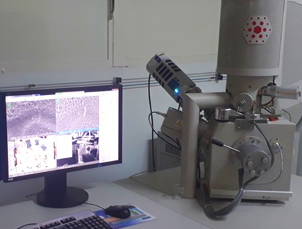
Scanning Electron Microscope - Quanta FEG 250 - FEI
● Everhart-Thornley detector (ETD): This detector is used for capturing secondary electron images, which are valuable for visualizing the sample’s topography and produce high resolution images.
● Circular backscatter detector (CBS): The CBS detector is utilized to obtain images based on atomic number contrast by detecting backscattered electrons.
● Large field detector (LFD): This detector is employed in Low Vacuum mode, allowing imaging of non-conductive and non-metalized samples.
● Environmental scanning electron microscopy (ESEM): The ESEM mode enables imaging in low vacuum conditions, making it suitable for samples with moderate water content (20-30%) or in a humid environment. This mode also facilitates in situ experiments.
● Scanning transmission electron microscopy (STEM): The STEM detector is used in transmission mode, enabling high-resolution imaging (up to 300,000x) of nanostructured samples.
● Energy dispersive spectrometer (EDS): The EDS detector, X-Max50 from Oxford, enables semi-quantitative elemental analysis of samples, as well as point analysis and elemental mapping. It offers high energy resolution and data acquisition speed.This microscope allows for high-resolution imaging even when operated at low electron acceleration voltages (typically up to 2 kV). This capability makes it possible to analyze samples that are sensitive to electron beams and samples with nanoscale dimensions, such as graphene oxide and cellulose nanocrystals. Samples not suitable for analysis include:
• Volatile substances
• High water content
• Samples that may contain solvents or oils
• Magnetic samples
• Corrosive samples
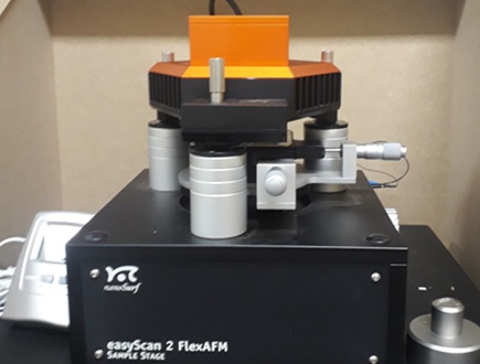
Atomic Force Microscope C3000 FlexAFM – Nanosurf
The atomic force microscope (AFM) doesn’t require extensive sample preparation, and analyses are conducted under ambient conditions, making it a complementary technique to scanning electron microscopy (SEM). The C3000 FlexAFM has a scanning limit of 100 µm and a Z-axis range of 10 µm. It is suitable for analyzing the surface of a wide range of materials, including those that cannot be analyzed by SEM, such as:
• Materials containing a high water content.
• Materials containing solvent and/or oil residues, including petroleum and asphaltenes.
• Magnetic materials.
The main operating modes are as follows:
• Non-contact/tapping: This is the standard mode of operation for AFM, which allows for obtaining information about the sample’s topography as well as phase contrast.
• Kelvin probe force microscopy (KPFM): The KPFM mode enables the acquisition of a surface potential map of the samples simultaneously with the topography image.
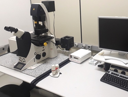
Laser Scanning Confocal Microscope – Leica TCS SP5
The confocal microscope enables the acquisition of optical sections of samples, forming well-defined images that can be used to generate a 3D image. Other available functions include the construction of emission spectra and real-time assays (time series).The available laser lines are:
Laser [Wave-length (nm)]
Argon [458, 476, 488, 496, 514]
He:Ne [543, 633]
It is suitable for biological samples, such as cell cultures or tissue sections, as well as for the analysis of materials (polymers, ceramics, and metals). Since it is a fluorescence microscope, in cases where the sample does not naturally fluoresce, a fluorophore with an absorption spectrum compatible with the available excitation wavelengths in the equipment should be added. Although it is not the conventional mode of operation, images of reflected light can also be obtained.The main experiments carried out on the Confocal Microscope are:
● Obtaining two-dimensional images
● Construction of three-dimensional images
● Labeling of specific structures through immunofluorescence
● Quantitative analysis
● Real-time experiments (time series)
● Obtaining fluorescence spectra
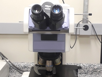
Optical microscope – Nikon E800
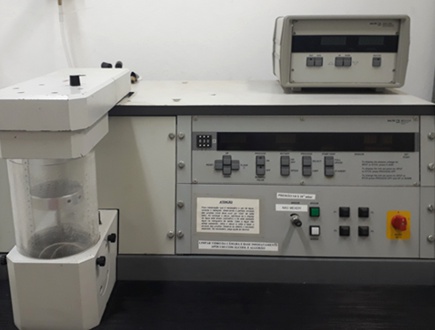
Sample Preparation Equipment: Metalizer MED020 – Baltec
The metal coater is used in sample preparation for scanning electron microscopy (SEM) analysis. Coating non-conductive samples is necessary to prevent charge build-up. The coating of samples can be done using:
•Sputtering:
• Au/Pd
• Iridium
•Carbon evaporation
Team

